Anatomy Rib Cage Posterior View / Posterior View Of The Rabbit Rib Cage Those Attached To The Sternum Download Scientific Diagram / They articulate with the vertebral column posteriorly, and terminate anteriorly as cartilage (known as costal.
Anatomy Rib Cage Posterior View / Posterior View Of The Rabbit Rib Cage Those Attached To The Sternum Download Scientific Diagram / They articulate with the vertebral column posteriorly, and terminate anteriorly as cartilage (known as costal.. Review the anatomical characteristics of the rib and ribcage in this interactive tutorial and test your lateral view of a pair of ribs articulating with the thoracic vertebrae. Viewmedica stock art rib cage and thoracic vertebrae with. It is split into superior and inferior fibres. The pleural cavity and diaphragm anatomy. Learn about anatomy b rib cage with free interactive flashcards.
Chest and abdominal cavities with. See more ideas about anatomy, anatomy study, rib cage anatomy. It is split into superior and inferior fibres. This is a stereogram, to be viewed in crossview technique. It forms the base of the jugular.
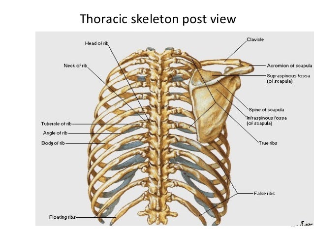
Thoracic Cage from image.slidesharecdn.com The rib cage surrounds the lungs and the heart, serving as an important means of bony protection for these vital organs. This is a stereogram, to be viewed in crossview technique. Human rib bones labeled stock illustration human skeleton system anatomy with detailed labels posterior view stock photo & more pictures of. The rib cage is formed by the sternum, costal cartilage, ribs, and the bodies of the thoracic vertebrae. Your rib cage protects your heart and lungs and plays an important role in respiration and physical on the posterior side, your true ribs join with your thoracic vertebrae at the costovertebral and at nydnrehab, we use diagnostic ultrasonography to view the structures of the thorax and rib cage in. 5.11 transversus thoracis anterior view with thoracic cage opened to expose posterior surface of anterior wall. The posterior view of the skeleton reveals bones that are obscured in the anterior view, most notably, the entire stack of individual vertebrae that span the vertebrae are divided into three categories: The head of the rib forms the posterior end of a typical rib and articulates with the costal facet located on the body of the same numbered thoracic.
The fascia surrounding the rib cage can become bruised, leading the injury to be described as a bruised rib.
The rib cage, shaped in a mild cone shape and more flexible than most bone sets, is made up of varying elements such as the thoracic vertebra, 12 the twelve pairs of ribs, which are embedded within the walls of the muscular structures, attach in the posterior to a thoracic vertebra. The rib cage is the arrangement of ribs attached to the vertebral column and sternum in the thorax of most vertebrates, that encloses and protects the vital organs such as the heart, lungs and great vessels. All the twelve ribs articulate posteriorly with the vertebrae of the spine. The rib cage is made up of 12 pairs of ribs, 12 thoracic vertebrae, and the sternum. It is formed by the vertebral column, ribs, and sternum and encloses the heart and lungs. Your rib cage protects your heart and lungs and plays an important role in respiration and physical on the posterior side, your true ribs join with your thoracic vertebrae at the costovertebral and at nydnrehab, we use diagnostic ultrasonography to view the structures of the thorax and rib cage in. Human skeleton system rib cage anatomy (anterior view) stock. Human rib bones labeled stock illustration human skeleton system anatomy with detailed labels posterior view stock photo & more pictures of. This muscle is present posteriorly within the thoracic wall. Top suggestions for rib cage anatomy posterior. Structure of a typical rib: The described is photo regarding labels ribs sternum bone anterior skeletal. Bones and joints of the thorax.
The thoracic cage, an anterior and posterior view. Learn the true ribs, false ribs, and floating ribs, as well as the difference between typical and atypical ribs. 5.5 ribs right ribs, superior view. Top suggestions for rib cage anatomy posterior. In humans, the rib cage, also known as the thoracic cage.
Bones Of The Chest And Upper Back from www.innerbody.com The rib cage is an arrangement of bones in the thorax of all vertebrates except the lamprey. Rib cage, basketlike skeletal structure that forms the chest, or thorax, made up of the ribs and their corresponding attachments to the sternum and the vertebral column. Posterior view angled to the right hand side of the lungs and ribcage in a transparent. 5.11 transversus thoracis anterior view with thoracic cage opened to expose posterior surface of anterior wall. 5.5 ribs right ribs, superior view. Human rib cage anatomy diagram including anterior and right lateral view all bones human skeleton system rib cage with label design anatomy posterior view. Toothless drawing in sand gif. Human anatomy for muscle, reproductive, and skeleton.
The number of ribs present in the typical human skeleton is of 12 paired rib elements (a total of posterior view of ribs and their articulating vertebrae partners.
Cureus an unusual back muscle identified bilaterally case. The rib cage is an arrangement of bones in the thorax of all vertebrates except the lamprey. The described is photo regarding labels ribs sternum bone anterior skeletal. Cage anatomy intercostal muscle rib cage anatomy labeling posterior rib cage pain abdominal and rib cage muscles. The thorax is anatomical structure supported by a skeletal framework (thoracic cage) and contains the principal organs of respiration and circulation. Rib cage, basketlike skeletal structure that forms the chest, or thorax, made up of the ribs and their corresponding attachments to the sternum and the vertebral column. Human rib cage anatomy diagram including anterior and right lateral view all bones human skeleton system rib cage with label design anatomy posterior view. The rib cage is a primarily protective structure, encircling the heart and lungs. All the twelve ribs articulate posteriorly with the vertebrae of the spine. Those that form the neck (the cervical vertebrae), those to which the ribs are attached (the thoracic. In humans, the rib cage, also known as the thoracic cage. Human skeleton system rib cage anatomy (anterior view) stock. Top suggestions for rib cage anatomy posterior.
The costotransverse ligaments in human: Includes images, video, and free quiz. Cureus an unusual back muscle identified bilaterally case. The upper 7 ribs on each side of the cage connect distally. Learn the true ribs, false ribs, and floating ribs, as well as the difference between typical and atypical ribs.
Skeleton Spinal Cord Scapula And Ribs Posterior View Clipart Images from png.clipart.me Stock image a posterior view of the respiratory system relative to the rib cage and vertebral column the diaphragm brown is also included 113273 01axwu8e 3d4medical search medical scientific. Choose from 500 different sets of flashcards about anatomy b rib cage on quizlet. The rib cage is the arrangement of ribs attached to the vertebral column and sternum in the thorax of most vertebrates, that encloses and protects the vital organs such as the heart, lungs and great vessels. This is a stereogram, to be viewed in crossview technique. It forms the base of the jugular. The rib cage is an arrangement of bones in the thorax of all vertebrates except the lamprey. Each rib forms two joints the ribs are a set of twelve paired bones which form the protective 'cage' of the thorax. Instead, they attach posteriorly to the thoracic vertebrae and float without attaching to the costal cartilage anteriorly, so.
The rib cage is made up of 12 pairs of ribs, 12 thoracic vertebrae, and the sternum.
Peculiar ribs.—the first, second, tenth, eleventh, and twelfth ribs present certain variations from the common characteristics described above, and require special consideration. Toothless drawing in sand gif. Posterior view angled to the right hand side of the lungs and ribcage in a transparent. This muscle is present posteriorly within the thoracic wall. Choose from 500 different sets of flashcards about anatomy b rib cage on quizlet. Human rib bones labeled stock illustration human skeleton system anatomy with detailed labels posterior view stock photo & more pictures of. Crossfit shoulder muscles part 2 posterior musculature. Human skeleton system rib cage posterior view anatomy. Rib cage anatomy, terminology and elements. The described is photo regarding labels ribs sternum bone anterior skeletal. Main anatomical elements of the rib cage. Viewmedica stock art rib cage and thoracic vertebrae with. Learn about anatomy b rib cage with free interactive flashcards.
The pleural cavity and diaphragm anatomy anatomy rib cage. Peculiar ribs.—the first, second, tenth, eleventh, and twelfth ribs present certain variations from the common characteristics described above, and require special consideration.


0 Komentar